The knee is made up of four bones. The femur or thighbone is the bone connecting the hip to the knee. The tibia or shinbone connects the knee to the ankle. The patella (kneecap) is the small bone in front of the knee and rides on the knee joint as the knee bends. The fibula is a shorter and thinner bone running parallel to the tibia on its outside. The joint acts like a hinge but with some rotation.
The knee is a synovial joint, which means it is lined by synovium. The synovium produces fluid lubricating and nourishing the inside of the joint. Articular cartilage is the smooth surfaces at the end of the femur and tibia. It is the damage to this surface which causes arthritis.
Femur
The femur (thighbone) is the largest and the strongest bone in the body. It is the weight bearing bone of the thigh. It provides attachment to most of the muscles of the knee.
Condyle
The two femoral condyles make up for the rounded end of the femur. Its smooth articular surface allows the femur to move easily over the tibial (shinbone) meniscus.
Tibia
The tibia (shinbone), the second largest bone in the body, is the weight bearing bone of the leg. The menisci incompletely cover the superior surface of the tibia where it articulates with the femur. The menisci act as shock absorbers, protecting the articular surface of the tibia as well as assisting in rotation of the knee.
Fibula
The fibula, although not a weight bearing bone, provides attachment sites for the Lateral collateral ligaments (LCL) and the biceps femoris tendon.
The articulation of the tibia and fibula also allows a slight degree of movement, providing an element of flexibility in response to the actions of muscles attaching to the fibula.
Patella
The patella (kneecap), attached to the quadriceps tendon above and the patellar ligament below, rests against the anterior articular surface of the lower end of the femur and protects the knee joint. The patella acts as a fulcrum for the quadriceps by holding the quadriceps tendon off the lower end of the femur.
Menisci
The medial and the lateral meniscus are thin C-shaped layers of fibrocartilage, incompletely covering the surface of the tibia where it articulates with the femur. The majority of the meniscus has no blood supply and for that reason, when damaged, the meniscus is unable to undergo the normal healing process that occurs in the rest of the body.
In addition, a meniscus begins to deteriorate with age, often developing degenerative tears. Typically, when the meniscus is damaged, the torn pieces begin to move in an abnormal fashion inside the joint.
The menisci act as shock absorbers protecting the articular surface of the tibia as well as assisting in rotation of the knee. As secondary stabilisers, the intact menisci interact with the stabilising function of the ligaments and are most effective when the surrounding ligaments are intact.
Anterior Cruciate Ligament
The anterior cruciate ligament (ACL) is the major stabilising ligament of the knee. The ACL is located in the centre of the knee joint and runs from the femur (thigh bone) to the tibia (shin bone), through the centre of the knee. The ACL prevents the femur from sliding backwards on the tibia (or the tibia sliding forwards on the femur). Together with the posterior cruciate ligament (PCL), ACL stabilises the knee in a rotational fashion. Thus, if one of these ligaments is significantly damaged, the knee will be unstable when planting the foot of the injured extremity and pivoting, causing the knee to buckle and give way.
Posterior Cruciate Ligament (PCL)
Much less research has been done on the posterior cruciate ligament (PCL) because it is injured far less often than the ACL. The PCL prevents the femur from moving too far forward over the tibia. The PCL is the knee’s basic stabiliser and is almost twice as strong as the ACL. It provides a central axis about which the knee rotates.
What is Arthritis?
Arthritis is a general term covering numerous conditions where the joint surface or cartilage wears out. The joint surface is covered by a smooth articular surface that allows pain free movement in the joint. This surface can wear out for a number of reasons; often the definite cause is not known. When the articular cartilage wears out the bone ends rub on one another and cause pain. This condition is referred to as Osteoarthritis or “wear and tear” arthritis as it occurs with aging and use. It is the most common type of arthritis.
Causes of Arthritis
There are numerous conditions that can cause arthritis but often the exact cause is never known. In general, but not always, it affects people as they get older (Osteoarthritis). Other causes include:
- Trauma (fracture)
- Increased stress such as overuse and overweight
- Infection of the bone
- Connective tissue disorders
- Inactive lifestyle and Obesity (overweight); Your weight is the single most important link between diet and arthritis as being overweight puts an additional burden on your hips, knees, ankles and feet.
- Inflammation (Rheumatoid arthritis)
Symptoms
Knee Arthritis causes pain and decreased mobility of the knee joint. In the arthritic knee there is an absent joint space that shows on X-ray. In the normal knee there is a normal joint space.
Arthritic knee
The cartilage lining is thinner than normal or completely absent. The degree of cartilage damage and inflammation varies with the type and stage of arthritis. The capsule of the arthritic knee is swollen. The joint space is narrowed and irregular in outline; this can be seen in an X-ray image. Bone spurs or excessive bone can also build up around the edges of the joint. The combinations of these factors make the arthritic knee stiff and limit activities due to pain or fatigue.
Diagnosis
Evaluating the source of knee pain is critical in determining your treatment options for relief of the pain. Knee pain should be evaluated by an Orthopaedic specialist for proper diagnosis and treatment. Your physician will perform the following:
- Medical History
- Physical Examination
Depending on what the history and exam reveal, your doctor may order medical tests to determine the cause of your knee pain and to rule out other conditions.
Diagnostic Studies may include:
- X-rays: X-rays are a form of electromagnetic radiation that is used to take pictures of bones. An X-ray can reveal if osteoarthritis from degenerative changes is causing your knee pain. The diagnosis of osteoarthritis is made on history, physical examination & X-rays.
- There is no blood test to diagnose Osteoarthritis (wear & tear arthritis).
Surgical Procedure
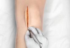
Total knee arthroplasty surgery is performed under sterile conditions in the operating theatre under spinal or general anaesthesia. You will be lying on your back and a tourniquet applied to your upper thigh to reduce blood loss. The surgeon makes an incision along the affected knee, exposing the knee joint. The surgeon first concentrates on the femur (thighbone).
The damaged portions of the femur are then cut at the appropriate angles using specialised jigs. The femoral component is attached to the end of the femur with or without bone cement. The damaged area of the tibia (shinbone) and the cartilage are cut or shaved. This removes the deformed part of the bone and bony growth, as well as allows for a smooth surface for which to attach the implants. The tibial component is secured to the end of the bone with bone cement or screws depending on a number of factors and on surgeon’s choice. The surgeon will place a plastic piece called an articular surface between the implants to assure a smooth gliding movement. This plastic insert will support the body’s weight and allow the femur to move over the tibia, similar to the original cartilage (meniscus). The femur and the tibia with the new components are put together to form the new knee joint. To make sure the patella (knee cap) glides smoothly over the new artificial knee: its rear surface is prepared to receive a plastic component. With all the new components, the knee joint is tested through its range of motion.
All excess cement will be removed. The entire joint will be irrigated or cleaned out with a sterile saline solution. The knee is then carefully closed and drains usually inserted and the knee dressed and bandaged.
Risks and Complications
- As with any major surgery there are potential risks involved. The decision to proceed with the surgery is made because the advantages of surgery outweigh the potential disadvantages.
- It is important that you are informed of these risks before the surgery takes place.
Complications can be medical (general) or local complications specific to the knee
Medical complications include those of the anaesthetic and your general wellbeing. Almost any medical condition can occur so this list is not complete. Complications include
- Allergic reactions to medications
- Blood loss requiring transfusion with its low risk of disease transmission
- Heart attacks, strokes, kidney failure, pneumonia, bladder infections
- Complications from nerve blocks such as infection or nerve damage
- Serious medical problems can lead to on-going health concerns, prolonged hospitalisation or rarely death.
Local complications
- Stiffness in the knee.
- Wound irritation or breakdown.
- Infection
- Blood clots (Deep Venous Thrombosis)
- Damage to nerves or blood vessels
- Wear
- Cosmetic Appearance
- Dislocation
- Patella problems
- Ligament injuries
- Fractures or breaks in the bone can occur during surgery or afterwards if you fall. To fix these, you may require surgery.